Home
/ Human Chest Muscles Diagram : Free Human Body Lesson Plan The Body S Systems Muscular System / 6 photos of the female chest muscles diagram.
Human Chest Muscles Diagram : Free Human Body Lesson Plan The Body S Systems Muscular System / 6 photos of the female chest muscles diagram.
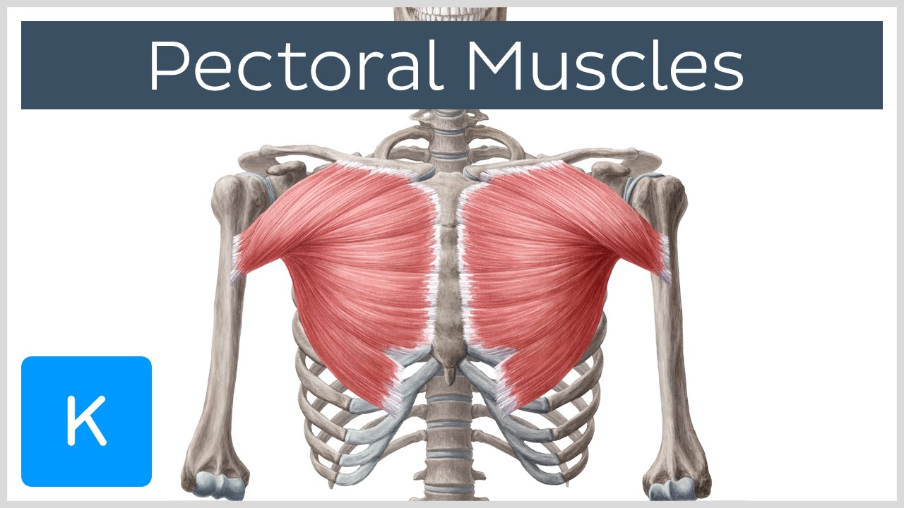
Human Chest Muscles Diagram : Free Human Body Lesson Plan The Body S Systems Muscular System / 6 photos of the female chest muscles diagram.. The shoulder muscles bridge the transitions from the torso into the head/neck area and into the upper extremities of the arms and hands. Human muscle system, the muscles of the human body that work the skeletal system, that are under voluntary control, and that are concerned with movement, posture, and balance. Free online quiz back and chest muscle diagram. Meet your pectoralis major and pectoralis. Primarily, there are three chest muscles involved.
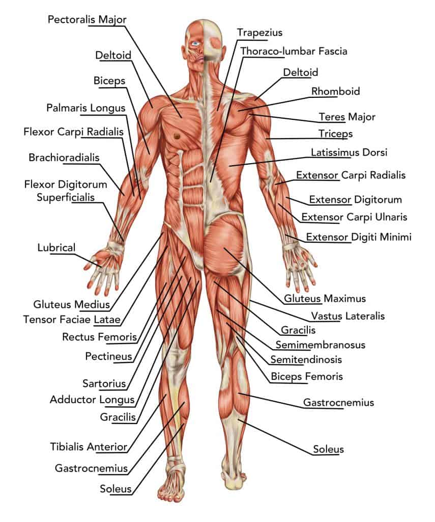
Related posts of chest muscles diagram anatomy muscle arm. Sternocleidomastoid muscle clavicle and ribs anatomy muscle anatomy chest sternocleidomastoid ribs anatomy chest muscles anatomy thorax rib muscles chest muscles chest anatomy illustration. Human body muscle system, the muscles of the human body that work the skeletal system, that are under voluntary control, and that are concerned with movement, posture, and balance. The dominant muscle in the upper chest is the pectoralis major. The bones of the appendicular skeleton provide support and flexibility at the joints and anchor the muscles that move the limbs.
Pectoral Muscles Area Innervation Function Human Anatomy Kenhub Youtube from i.ytimg.com The shoulder muscles bridge the transitions from the torso into the head/neck area and into the uppe. Our latest youtube film is ready to run. The average human heart weighs between 6 and 11 ounces. Meet your pectoralis major and pectoralis. See human chest anatomy stock video clips. Muscle charts of the human body for your reference value these charts show the major superficial and deep muscles of the human body. Anatomy of the rib cage diagram. See the anatomy of muscle movement in 3d.
The back has a total of 40 muscles.
Each of these muscles is a discrete organ constructed of skeletal muscle tissue, blood vessels, tendons, and nerves. Likewise, there are muscles in other parts of the body that help support and move the spine. Pectoral muscles are most predominantly associated with movement of the shoulders and arms. The muscles of the chest and upper back occupy the thoracic region of the body inferior to the neck and superior to the abdominal region and include the muscles of the shoulders. Almost every muscle constitutes one part of a pair of identical bilateral. Each of these muscles is a discrete organ constructed of skeletal muscle tissue, blood vessels, tendons, and nerves. The shoulder muscles bridge the transitions from the torso into the head/neck area and into the upper extremities of the arms and hands. Primarily, there are three chest muscles involved. Related posts of chest muscles diagram anatomy muscle arm. Our latest youtube film is ready to run. See human chest anatomy stock video clips. Anatomy of the rib cage diagram. Below is a diagram showing the chest muscles depicting where the different exercises target.
Human chest muscles diagram : Anatomy of the rib cage diagram. Pectoral muscles are most predominantly associated with movement of the shoulders and arms. Major muscles back muscles shoulder muscles supraspinatus muscle back workout routine sternocleidomastoid muscle muscle diagram body diagram latissimus dorsi. It consists of the manubrium on its uppermost end, its body in the intermediate region and the small xiphoid process on its lowermost end.
The Complete Guide To Upper Body Muscles For Beginners Empower Your Wellness from empoweryourwellness.online Broadly considered, human muscle—like the muscles of all vertebrates—is often divided into striated muscle, smooth muscle, and cardiac muscle. The bones of the appendicular skeleton provide support and flexibility at the joints and anchor the muscles that move the limbs. Chest muscles, chest muscle diagram. Related posts of chest muscles diagram anatomy muscle arm. For that reason, and because of the dexterity of the shoulder joint itself, the musculature of the shoulder is. The bones of the skeletal system act as attachment points for the skeletal muscles of the body. The muscles of the chest and upper back occupy the thoracic region of the body inferior to the neck and superior to the abdominal region and include the muscles of the shoulders. It extends from the position of the diaphragm to the clavicle or the collar bone.
It extends from the position of the diaphragm to the clavicle or the collar bone.
The chest is the area of origin for many of the body's systems as it houses organs such as the heart, esophagus, trachea, lungs, and thoracic diaphragm. Diagram of a long bone anatomy 12 photos of the diagram of a long bone anatomy bone function, describe the structure of a bone, diagram compact bone, diagram femur, diagram osteon, structure of bones, what does spongy bone do, human anatomy, bone function, describe the structure of a bone, diagram compact bone, diagram femur, diagram … The shoulder muscles bridge the transitions from the torso into the head/neck area and into the upper extremities of the arms and hands. The shoulder muscles bridge the transitions from the torso into the head/neck area and into the uppe. Alles rund um kostüme & verkleiden. Free online quiz back and chest muscle diagram. Human chest muscles diagram : Each of these muscles is a discrete organ constructed of skeletal muscle tissue, blood vessels, tendons, and nerves. Human muscle system, the muscles of the human body that work the skeletal system, that are under voluntary control, and that are concerned with movement, posture, and balance. Broadly considered, human muscle—like the muscles of all vertebrates—is often divided into striated muscle, smooth muscle, and cardiac muscle. The dominant muscle in the upper chest is the pectoralis major. The sternum is a nearly flat rigid bone in the middle of human chest. The bones of the skeletal system act as attachment points for the skeletal muscles of the body.
The chest muscles are made up of the pectoralis major and, underneath that, the pectoralis minor. The average human heart weighs between 6 and 11 ounces. Extends across body nd separates chest cavity from abdomen cavity. See more ideas about muscular system, anatomy, muscle anatomy. Muscles allow a person to move, speak, and chew.
Anatomy Drawing Conor Power Shoulder Anatomy Chest Muscles Muscle Diagram from i.pinimg.com Pectoral muscles are most predominantly associated with movement of the shoulders and arms. The sternum is a nearly flat rigid bone in the middle of human chest. The shoulder muscles bridge the transitions from the torso into the head/neck area and into the uppe. Likewise, there are muscles in other parts of the body that help support and move the spine. It extends from the position of the diaphragm to the clavicle or the collar bone. The muscular system is responsible for the movement of the human body. Human chest muscles diagram : Below you'll see diagrams along with the names of the back muscles that may be the cause of your pain.
How many muscles are in the back?
Our latest youtube film is ready to run. There are around 650 skeletal muscles within the. The sternum is a nearly flat rigid bone in the middle of human chest. Human body muscle system, the muscles of the human body that work the skeletal system, that are under voluntary control, and that are concerned with movement, posture, and balance. The average human heart weighs between 6 and 11 ounces. Anatomy of the rib cage diagram. 6 photos of the female chest muscles diagram. Related posts of chest muscles diagram anatomy muscle arm. The bones of the skeletal system act as attachment points for the skeletal muscles of the body. Human chest muscles diagram : The major muscle in the chest is the pectoralis major. A woman's chest — like the rest of her body — is covered with skin that has two layers. The dominant muscle in the upper chest is the pectoralis major.
